
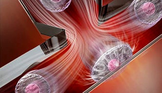

Microfluidic single-cell smasher: A novel inertial microfluidic cell stretcher (iMCS) capable of characterizing and classifying large populations of single-cell deformability near real-time in a fully automated manner.
A team including researchers at Rensselaer has developed an innovative approach to measuring cellular mechanical properties (i.e., cell stiffness) that is part of an emerging label-free (i.e, no histology dyes or immunolabeling) biophysical marker that can be used for the identification of cell diseases and cellular states. The research is important, since it can be used for rapid cancer diagnosis and rapid drug screening, as well as the development of personalized medicine.
As reported in a paper recently published in Small, researchers at Rensselaer have developed a process that measures thousands of single-cell stiffness using a novel inertial microfluidic cell stretcher (iMCS) in a fully automated and high-throughput manner for cell state identification.

Aram Chung
According to the researchers―lead authors at Rensselaer, along with a colleague from the University at Albany―it is a well-known fact that malignant cancer cells are softer (more deformable) than regular healthy cells. As another example, red blood cells (RBCs) from a malaria-infected patient are more rigid than a healthy person’s RBCs. “These observations suggest that measuring cellular mechanical properties is a crucial task, and this principle has been widely applied in biophysical studies, cancer diagnosis, and drug discovery,” said Aram Chung, an assistant professor in the Department of Mechanical, Aerospace, and Nuclear Engineering.
“The challenge is that scanning thousands of single-cell stiffness in a robust, rapid, and automated manner is extremely difficult,” Chung said. “In pursuing this research, the challenges had to be addressed through innovations from multiple fields, and a strong collaboration among cancer biologists and mechanical and biomedical engineers, to enable the research. Here, we report a novel microfluidic device characterizing more than 6,000 cells’ mechanical properties from sample injection to data analysis within one minute.”
Chung noted that various applications are expected from their development. First, iMCS shows great potential to be implemented clinically for rapid cancer diagnosis by processing unknown patient samples. For example, ascites or pleural effusion samples can be directly collected from patients and processed through iMCS, where immediate cell deformability results can be obtained, which could potentially guide physicians in a timely manner. Furthermore, the research team’s platform can be expanded for rapid drug screening and personalized medicine. Instead of waiting for days to obtain the efficacy of drug treatment on specific samples at different conditions, iMCS offers rapid cell response characterization under various external stimuli, providing a possibility for processing a large number of samples for various drug testing applications.
“As the first author of the paper, my role was focused on engineering a microfluidic platform that can actually be used for medical and biophysical research. To enable this, I spent much of my time working to design a simple iMCS to use while not sacrificing sophisticated functionalities for precise single-cell mechanical characterization,” said Yanxiang Deng, a graduate student who also works in Professor Chung’s Bio-Optofluidics Laboratory.
The Bio-Optofluidics Laboratory is a research group in the Department of Mechanical, Aerospace, and Nuclear Engineering that is devoted to research on investigating microscale flows at finite Reynolds numbers. Based on a series of fundamental studies of inertial fluid behaviors in microscale and their integration with other modalities, the lab is developing a new class of practical high-throughput microfluidic platforms for biomedicine and manufacturing.
At Rensselaer, Deng’s area of research focuses on studying inertial microfluidic physics to develop novel microfluidic microsystems for quantitative single-cell analysis. As his next step, Deng is working on creating a novel deformability-based cell sorter enabling downstream molecular profiling to understand the relation between deformability and cancer migration through a new collaboration with the Albany Medical Center.
“Besides engineering an innovative microfluidic system for real-time cell mechanical screening, our automation effort should be highlighted,” Chung said. “Currently, many microfluidic developments have not been substantially adopted by biological or medical personnel. This is largely because they are hard to operate without extensive training or/and relevant backgrounds. Here, we have enabled a single-command-based fully automated operation where the process runs without any human intervention, realizing a user-friendly and robust operation.” Chung mentioned that without simplicity and robustness, microfluidic technologies would not make practical impacts, and he continuously emphasizes this to his research group.