
Contributors: Regina Stracqualursi and Torie Wells
According to the American Cancer Society, more than 1.7 million new cancer cases are expected to be diagnosed in the U.S. this year, and over 600,000 individuals are expected to die of the disease. As the second leading cause of death in the nation, cancer has affected most people in one way or another — whether it be a personal diagnosis or experience supporting a loved one who is battling the disease.
Part of what makes some cancers so difficult to treat is the inability to closely monitor how individual cells respond to drug treatments. If researchers and scientists were able to see a drug’s effect on cancer cells in realtime, they would be able to tailor treatments accordingly.
Engineers at Rensselaer Polytechnic Institute have partnered with biologists from Albany Medical College and engineers from MARS Bioimaging Ltd to tackle this challenge. As part of a five-year project supported by the National Institutes of Health, the team is working to develop imaging technology that could aid in the treatment of breast cancer.
The team, purposefully multidisciplinary, is tackling the problem from many angles, combining optical imaging and X-ray imaging technologies to provide not only a deeper look at cancer cells but also the ability to look at cancer in living systems.
Ge Wang, director of the Biomedical Imaging Center at Rensselaer, and his research team worked with MARS Bioimaging to develop a new type of X-ray imaging that allows a user to see images in color, creating a clear visual differentiation between soft tissue, bone, water, and treatment drugs.
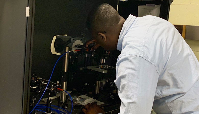
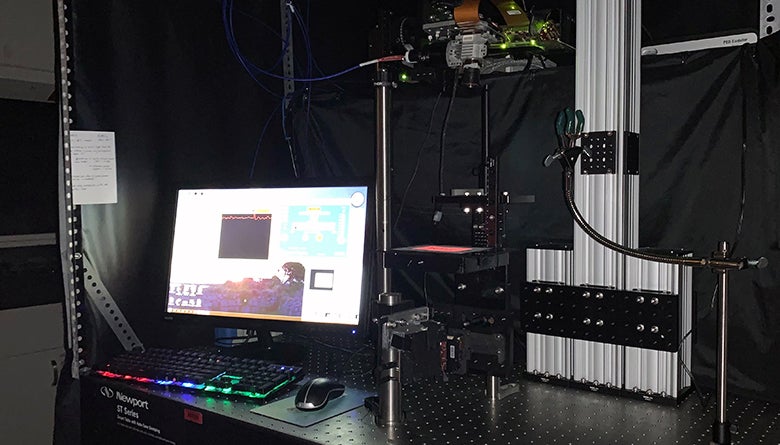
Xavier Intes, professor of biomedical engineering at Rensselaer and co-director of the Biomedical Imaging Center, worked with his research team and Albany Medical College to develop optical imaging technology that utilizes lasers to observe the delivery of drugs to a cancer cell.
Using these two technologies together, the team has captured clearer and more detailed images than was previously possible.
The combination of optical and X-ray imaging has the potential to allow those researching and developing treatments for cancer to monitor the interaction between drugs and cancer cells closer than ever before. It could also give doctors the ability to better tailor treatments based on how individual cancer patients respond to drug treatments.
To see some of the imaging technology in action, check out the interviews featuring the work on Spectrum News and News10 ABC in Albany, New York.
Pictured Above:
The lab of Professor Xavier Intes.